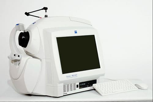
Carl Zeiss Cirrus 500 Spectral Domain OCT HD with Windows, Power Table, Printer and Full Warranty. Excellent condition and completely refurbished by our Zeiss trained technician.
Thank you for viewing our listing! This Cirrus 500 has been completely serviced and refurbished by our trained staff of technicians. This unit comes with Windows Ultimate, new printer, power table, and all peripherals. The system software has been upgraded and several optional software licenses are active. See photos!
The Cirrus 500 is the current model offered by Zeiss, and they are very difficult to find on the used market. This OCT can be shipped almost anywhere in the world. Cirrus is the world’s best-selling spectral domain OCT system!
Visualization at the speed of Cirrus
Analyzing a single pathology from multiple views provides comprehensive insight and analysis of the clinical situation. How this helps you: Spot small areas of pathology. Tightly spaced B-scans, (either 30 or 47 μm apart), in the cube ensure that small areas of pathology are imaged. For reference, a human hair is about 40-120 μm in diameter. Visualize the fovea. Scans that are spaced further apart than in the CIRRUS cube may miss the central fovea. Fuel for analysis. Millions of data points from the cube are fed into the Zeiss proprietary algorithms for accurate segmentation, reproducible measurements and registration for change analysis. Take the pressure off the operator. As long as the scan is placed in the vicinity of the fovea or optic nerve, the software automatically centers the measurements after the capture. See the tissue from different perspectives. View the cube data from all angles, with 3D rendering, OCT fundus images and Advanced Visualization™. Future-ready. Previously captured CIRRUS cubes can be analyzed using new analyses.
Tracking at the speed of Cirrus
FastTrac™ reduces eye motion artifacts without sacrificing patient throughput with a proprietary scan acquisition strategy, high speed 20 Hz LSO camera, and single-pass alignment scanning
With FastTrac, scan at the highest resolution at the same location at each visit.
Assessment at the speed of Cirrus
Measurement centering with FoveaFinder and AutoCenter
On the macula, the unique FoveaFinder technology ensures the ETDRS and ganglion cell plus inner plexiform layer measurement frameworks are centered on the fovea.
AutoCenter™ function automatically centers the 3.4 mm diameter peripapillary RNFL calculation circle around the disc for precise placement and repeatable registration. The placement of the circle is not operator dependent. Accuracy, registration and reproducibility are assured.
Specifications
OCT Imaging
Methodology: Spectral domain OCT
Optical source: Superluminescent diode (SLD), 840 nm
Scan speed: 27K- 68K A-scans per second
A-scan: 2.0 mm (in tissue), 1024
Axial resolution: 5 μm (in tissue)
Transverse resolution: 15 μm (in tissue)
Fundus Imaging
Methodology: Line scanning opthalmoscope (LSO)
Live fundus image: During alignment and during OCT scan
Optical source: Superluminescent diode (SLD), 750 nm
Field of view: 36 degrees W x 30 degrees H
Frame rate: > 20 Hz
Transverse resolution: 25 μm (in tissue)
Iris Imaging
Methodology: CCD camera
Resolution: 1280 x 1024
More photos available! Just ask.
octs
Zeiss Cirrus OCT Model 4000
Excellent refurbished condition. Our Zeiss Cirrus 4000 has been completely tested by our Zeiss trained technician. Includes full warranty.
This Cirrus 4000 is in excellent, lightly used condition and includes the original accessories, new printer, new peripherals and power table. Many of the optional licenses are also already active. See photos!
Each system we sell is fully serviced in-house by our trained technicians prior to sale. The CPUs are refurbished down to the component level and we guarantee everything we sell.
The Dual Core Cirrus 4000 is an excellent value for those looking for a Spectral Domain HD OCT at a reduced price. We refurbish these at component level and include a full warranty. We also have newer 4000 models, 500 and 5000.
Please watch our video below to learn more about our company, our great staff and our headquarters located in Tampa, Florida, USA.
Features
Built on 10 years’ experience at the vanguard of innovation, Carl Zeiss Meditec OCT technology has become the recognized standard of care. Now, Cirrus HD-OCT offers another leap forward with a superior platform that delivers unprecedented imaging details for clinical decision making.
ZEISS optics provide superior visualization of anatomical details across a wider range of patients.
Robust engineering with premium components ensures consistent precision performance.
Unique HD layer maps and images highlight clinically relevant details for identification and monitoring of specific diseases – all at a glance.
The powerful Cirrus HD-OCT scan engine delivers superior image data. The new HD Enhanced Raster Scan leverages this power to produce images with outstanding detail while maintaining patient throughput. The proprietary Selective Pixel Profiling™ technology enhances anatomical features while reducing image noise.
Cirrus HD-OCT enables repeatable visualization of clinically relevant anatomy with exact correlation between the OCT scan and the fundus image. Comprehensive navigational tools ensure efficient and simple operation.
Designed for efficiency
Small footprint and integrated design are ideal for crowded or busy practice
90-degree orientation facilitates observation of patient throughout exam
Advanced optics aid in the examination of patients with cataracts
Dilation is not required even for pupils as small as 2.5 mm
Mouse Driven Alignment delivers superior image capture and analysis in just a few clicks, resulting in reduced chair time for the patient
Auto Patient Recall assures patient position and instrument setting are repeated from previous visit
Technical Data
OCT Scanning
-Axial resolution: 5 μm (in tissue)
-Transverse resolution: 15 μm (in tissue)
-Scan speed: 27,000 A-scans per second
-A-scan depth: 2.0 mm (in tissue), 1024 points
-Optical source: superluminescent diode (SLD), 840 nm
Fundus Imaging
-Line scanning ophthalmoscope (LSO)
-Live during scanning
-Transverse resolution: 25 μ (in tissue)
-Optical source: superluminescent diode (SLD), 750 nm
-Field of view: 36° x 30°
Scan Patterns
-Macular Cube 200 x 200 Combo: 200 horizontal scan lines comprised of 200 A-scans
-Macular Cube 512 x 128 Combo: 128 horizontal scan lines comprised of 512 A-scans
-5 Line Raster: 4096 A-scans per B-Scan (adjustable length, spacing and orientation)
Focus Adjustment Range
−20D to +20D (diopters)
Fixation
-Internal and external
Computer
-Windows OS
-Internal storage: > 80,000 scans
-CD-RW, DVD-ROM drive
-Integrated 15″ color flat panel display
Pupil Size Requirement
-≤ 2.0 mm (≥ 3.0 mm optimal for LSO)
Dimensions/Weight (Instrument Only)
-25.6 L x 17.3 W x 20.9 H (in); 65 L x 44 W x 53 H (cm)
-83 lbs; 37.6 kg
Electrical
-100-120V~, 50/60Hz, 5A 220-240V~, 50/60Hz, 2.5A
More photos available! Just ask.
Keep in mind we are not brokers. We are a full sales and service center. Each laser and piece of equipment we sell is hand picked, inspected and tested by our technicians and engineers to ensure your order arrives in excellent condition both cosmetically and mechanically. Buy with confidence from Laser Locators!
Zeiss Cirrus OCT Model 400
Thank you for viewing our listing! This Cirrus 400 has been completely serviced and refurbished by our trained staff of technicians. This unit comes with software, printer, power table and all peripherals as shown.
The world’s best-selling spectral domain OCT system is now offered in Model 400. What’s unique about Model 400? Focused on the essential core OCT functionality, Model 400 is designed with the smaller budget in mind. Live OCT Fundus technology provides the fundus image using the OCT scanner only, rather than an additional line scanning ophthalmoscope (LSO).
The 400 offers glaucoma and retina analyses and is capable of anterior segment imaging just like the 4000. The dense data cubes are interchangeable between model 400 and model 4000 systems. Cirrus HD-OCT models also share the same modern integrated design, ease of use, and small footprint.
Features
- Macular Thickness Analysis
- RNFL Thickness Analysis
- Normative Database – RNFL
- Advanced Visualization and 3D Display
- Anterior Segment Imaging
- 5-line Raster Scan
- Compatible with Cirrus Review Software (license sold separately)
Includes
- Fundus imaging
- Scanning system Live OCT Fundus Technology
- Field of view 36 x 22 degrees
- OCT imaging
- Methodology Spectral domain
- Scan speed 27,000 A-scans / sec
- A-scan depth 2.0 mm (in tissue)
- Axial resolution 5 µm (in tissue)
- Transverse resolution 15 µm (in tissue)
Computer
- Processor Intel Pentium Core 2 Duo, Win XP OS
- Memory 2GB
- Hard Drive 750GB
Physical
- Size 26L x 17W x 21H (in)
- Table footprint 39L x 22W (in)
Zeiss Vistante OCT
Key Features
- Provides excellent high-resolution images of the anterior segment of the eye
- Uses low-coherence interferometry to produce cross-sectional tomographs of the cornea and anterior chamber
- A non-contact instrument that captures its images quickly and with little technician effort
- Extremely useful in determining residual stromal bed for the purposes of LASIK enhancement and monitoring cases of endothelial keratoplasty
Technical Specifications
- Illumination laser source long wavelength 1,310 nm superluminescent LED Scan types
- Anterior segment — Single, dual and quad line scans (16 mm x 6 mm) Adjustable in 1-degree increments 256 A scans per line sampling 0.125 second per line acquisition time
- Pachymetry map — Adjustable 8-line scan pattern (10 mm x 3 mm) Regional map with maximum, minimum and average values 128 A scans per line sampling 0.5 second total acquisition time
- High-resolution corneal scans Adjustable in 1-degree increments (10 mm x 3 mm) 512 A scans per line sampling 0.25 seconds per line acquisition time
- Optical resolution Axial: 18 μm Transverse (center): 60 μm
- Ametropia correction: -35 to +20 diopters
- Fixation target: Internal or external
- Display: Integrated 15-inch flat-panel display
- Computer specs: Windows / 3.0 GHz Pentium IV / 1 GB memory
Dimensions/Weight
- 48.5 cm H x 43.8 cm W x 63.2 cm D; 34.5 kg
- (19.1 inch H x 17.2 inch W x 24.9 inch D) 76.1 lb
OCTs
OCTs
Each optical coherence tomography system offers varying features from incorporated high resolution fundus cameras to analytical tools. We carry OCTs such as the Zeiss Cirrus 4000, Zeiss Stratus OCT 3000, Heidelberg HRT-II, Heidelberg HRT-III, Heidelberg Spectralis, Topcon 3D OCT-2000 and Optovue RTvue.