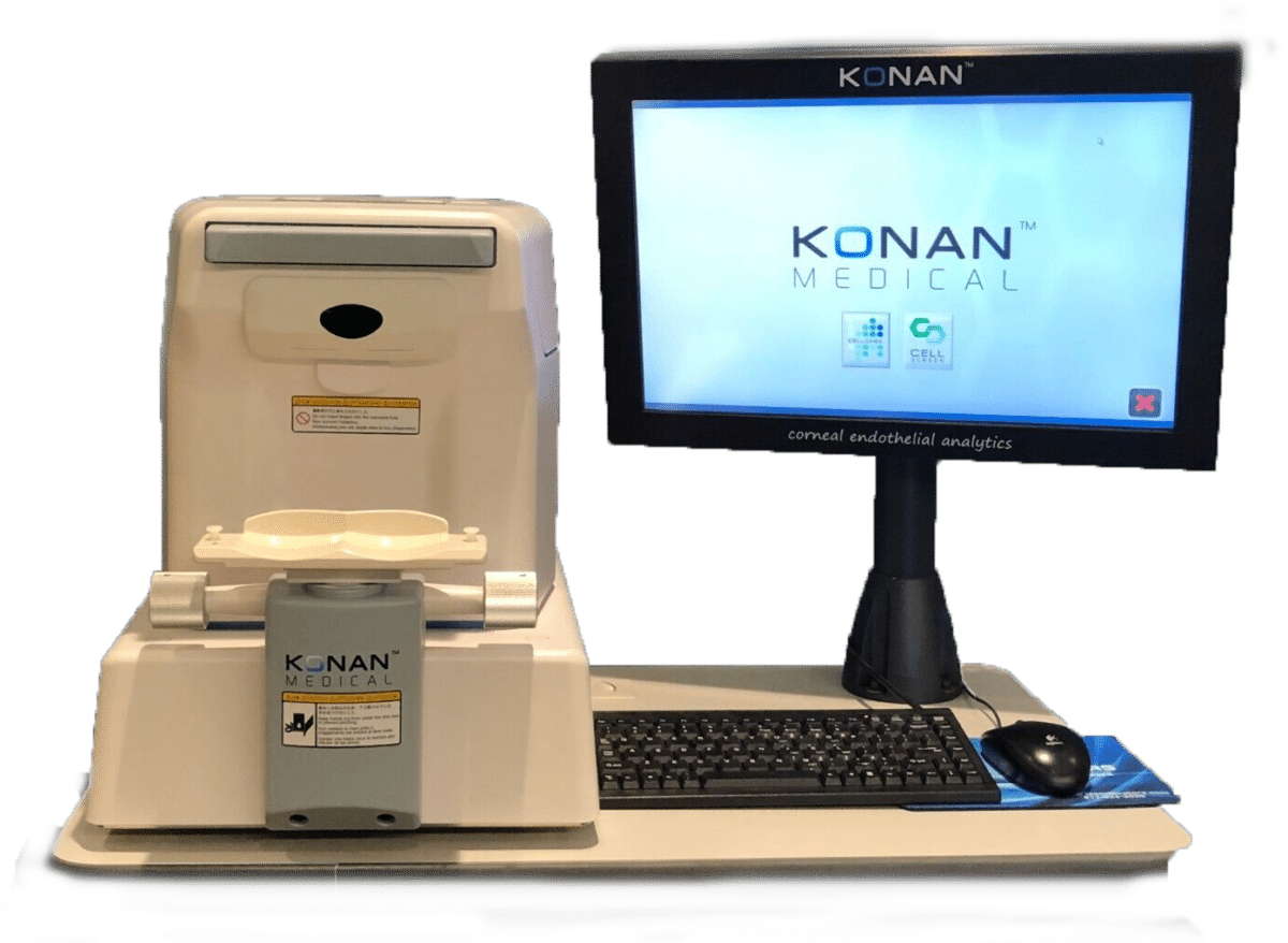
Konan sets the standard for specular microscopy with strong clinical evidence, ease of use, and patented analysis methods that can reliably assess even problematic endothelium.
- CellChek Specular Microscope
- Cornea Fundamentals
- Clinical Resources
- Testimonials
- Specifications
- Support FAQs
- Customer Service
Fully Automated
Konan’s CellChek specular microscope features auto-align, auto-focus, auto-caputure, auto-analysis, and auto-pachymetry for one-button ease of use. But this is where others end. Konan additionally includes semi-automated methods (see Analysis Methods section) to make robust use of minim numbers of observable cells with advanced disease state corneas.
Trends Analysis – Only from Konan
Konan’s patented capture method acquires data samples that include position data which can allow accurate re-assessment of same specular data sample areas to trend cellular statistics over time. Trends analysis is critical to understanding your chosen treatment responses and progression or arrest of disease.
FDA 510(k) cleared under “Specular Microscopes” Product Code NQE. Why accept anything less than the assurance of the FDA’s review for both imaging and assessment of the corneal endothelial cell layer, morphology of endothelial cells, and corneal pachymetry.
Specular Microscopes
For practices that want greater detail on a cellular level than biomicroscopy provides, browse Laser Locators’ refurbished specular microscopes. We offer several model options including the Konan Noncon Robo and Topcon SP-2000P to meet your specular microscope needs.
Konan
Category:Specular Microscopes
Konan
Category:Specular Microscopes
Konan
Category:Specular Microscopes
Topcon
Category:Specular Microscopes
Dual Measurement System:
Corneal Endothelium Measurement
Corneal Thickness Measurement
Alignment System:
This innovative auto alignment and auto capture system ensures ease of operation and reliable results.
Built-In Fixation Targets:
Corneal thickness and endothelium images can obtained of the central and four peripheral areas.
Peripheral fixation is established at the 12,2,6 and 10 o’clock positions.
Different colored LED’s aid in patient compliance.
Non-Contact:
The SP-2000P captures the image of the endothelium cells and calculates cornea thickness by a unique method that does not require touching the cornea. This patented procedure eliminates the risk of transmitting infectious disease and reduces potential physical injury to the eye. Patient comfort is increased. Because of greater patient cooperation, the examination time is greatly reduced.
Due to the non-contact operation, image acquisition can be obtained through contact lenses which allows the cell condition to be observed while fitting contacts.
Light Source For observation : Near infrared Light
For photography : Max. 60W sec. Xenon Flash;
Low & High adjustments
Photographic Area 0.2 × 0.5mm
Working Distance 25mm
Cell Counting Grid 1000 / 1500 / 2000 / 2500 / 3000 cell/mm
Image Memory 3 images for each eye (Total 6 Images)
Automatic Shut-off Standard
Power Voltage AC100, 120, 220, 240V 50 / 60 Hz
Power Consumption 110VA
Base Travel 40mm (back & forth), 88mm (right& left)
Head Travel 10mm (back & forth), 10mm (right& left)
30 mm (motorized vertical movement)
Chinrest Vertical Travel 60mm
Dimensions: 274(W) × 485(D) × 410(H)mm
Weight: 20kg