Zeiss Cirrus OCT Guide
10/08/2020
Zeiss Cirrus OCT Guide
What is the Zeiss Cirrus OCT?
Optical Coherence Tomography (OCT) is a non-invasive imaging technique used in ophthalmology to obtain high-resolution cross-sectional images of the retina and anterior segment. Zeiss Cirrus OCT functions like optical ultrasound, allowing eye care professionals to assess glaucoma, macular degeneration, diabetic macular edema, and other retinal diseases with micrometer precision.
How Do Eye Doctors Use Zeiss Cirrus OCT?
Ophthalmologists and optometrists use OCT to analyze retinal layers, optic nerve fiber thickness, and vascular health. OCT Angiography (OCTA) provides detailed blood flow imaging in the retina without the need for contrast dye. Glaucoma specialists utilize OCT to measure axonal thickness and optic nerve changes over time.
Evolution of Zeiss Cirrus OCT Technology: From Stratus to Cirrus
Early OCT Models
1996: First OCT instruments introduced, but adoption was slow.
2002: Stratus OCT by Zeiss became the first widely accepted OCT, scanning at 400 A-scans per second.
2004: Over 10 million OCT scans performed worldwide, proving its clinical necessity.
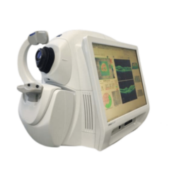
Current OCT Systems
Today, Cirrus OCT by Zeiss is the most widely used OCT system, known for its faster scanning, higher resolution, and ease of use.
Zeiss Cirrus OCT Series: Comparing the 400, 4000, 500, and 5000 Models
Cirrus 400 – Core OCT Capabilities at an Affordable Price
- Spectral-Domain OCT with high-resolution retinal imaging.
- Glaucoma and retina analysis included.
- Limited anterior segment imaging.
- Compact design with built-in Windows 10 for seamless operation.
Cirrus 4000 – Faster and More Advanced Imaging
- 27,000 A-scans per second for detailed retinal visualization.
- High-density data cubes allow 3D viewing of retinal structures.
- No need for external workstations – Windows 10 is built into the system.
- More efficient scan acquisition compared to Stratus OCT.
Cirrus HD-OCT 500 & 5000 – Next-Generation OCT Technology
- Zeiss Smart OCT with AI-assisted analytics.
- FastTrac™ retina tracking technology (exclusive to Cirrus 5000).
- 3D microvascular imaging for enhanced pathology assessment.
- Real-time motion correction for accurate scans over multiple visits.
Why Choose Cirrus HD-OCT for Your Practice?
- Comprehensive data analysis – Multi-layer retinal scans with 3D visualization.
- User-friendly operation – Designed for fast, intuitive use.
- Built-in Windows 10 – Eliminates the need for external hardware.
- Auto-tracking & AI-powered diagnostics – Increases accuracy and workflow efficiency.
Need help choosing the right OCT for your clinic?
Contact us today for a consultation.
Trusted by ophthalmologists worldwide.