Essential Equipment of Ophthalmology

Posted by Laser Locators
Slit Lamp
A slit lamp is a device consisting of a high-intensity light source that can shine a thin beam of light into the patient’s eye. The lamp allows for examining the human eye’s anterior and posterior segments, including the eyelid, conjunctiva, sclera, natural crystalline lens, iris, and cornea. The slit-lamp examination provides a magnified view of the eye in detail, enabling diagnoses to be made for various eye conditions. A smaller, hand-held lens is used to examine the retina.
Goldmann tonometer
Considered the Gold standard according to the AAO, a Goldmann is attached to a slit lamp and provides accurate and reproducible readings. Using a prism measures the force needed to flatten a 3.06mm diameter circle of the central cornea.
Non-Contact tonometer (NCT)
Also referred to as an “air puff” tonometer, it is a diagnostic tool used by eye care professionals to measure the intraocular pressure (IOP) inside a patient’s eyes. A non-contact tonometer uses a small puff of air to measure the eyes pressure. An industry term for this is also called a “puff test.”
Autorefractor/Keratometer (ARK)
A computer-based instrument is used to help determine the eyeglasses prescription. The Keratometer is a device used to determine the curve of the cornea. These measurements are typically taken on patients who are being fitted for contact lenses or who may have corneal problems.
Optical coherence tomography (OCT)
An instrument that takes transpupillary images of the retina to assist in diagnosing and treating retinal diseases.
Visual Field Machine (HFA)
The visual field is how wide an eye can see when focused on a central point. Visual field testing is one way an ophthalmologist measures how much vision there is in either eye or vision loss over time.
Visual field testing can determine if there are blind spots (scotoma) in a patient’s vision and where they are. A scotoma’s size and shape can indicate how eye disease, or a brain disorder affects one’s vision. For example, this test shows any possible side (peripheral) vision loss from this disease with glaucoma.
Ophthalmologists also use visual field tests to assess how vision may be limited by eyelid problems such as ptosis and droopy eyelids.
Fundus Camera
A fundus camera is a low-power microscope attached to a digital camera used to examine structures such as the retina, optic disc, and lens.
Topographer (Topo)
Corneal topography is a computer-assisted diagnostic tool that creates a 3D image of the cornea’s surface curve. The cornea is responsible for approximately 70% of the focusing power of the eye. An eye with good vision has an evenly curved cornea, but if the cornea is too flat or too steep, the vision will be less than perfect. The most significant advantage of corneal topography is the ability to detect rare conditions invisible to conventional testing.
Biometer/A-Scan (Ultrasound)
A-scan ultrasound biometry, commonly known as an A-scan, is a diagnostic test used in ophthalmology. An A-scan provides data on the eye’s length, which is used to screen for sight disorders. One of the A-scan uses in determining the eye’s size for calculating intraocular lens power for cataract surgery.
Phacoemulsification Machine (Phaco)
Phacoemulsification is a modern cataract procedure in which the eye’s lens is emulsified with an ultrasonic handpiece and aspirated from the eye. Fluids are replaced with irrigation of salt solution to maintain the anterior chamber. This procedure is also now performed with a femtosecond laser.
Green (532nm), Red(810nm), and Yellow(577nm) lasers
The most commonly employed wavelength in vitreoretinal practice is 532 nm green, widely referred to as an Argon Laser, and is used for treating retinal pathologies with pan-retinal photocoagulation. The 577 nm yellow laser is slightly absorbed by xanthophylls and well absorbed by oxygenated hemoglobin, making it the laser of choice for lesions near the macula. Good results with dye lasers operating at this wavelength have been reported.1 Krypton lasers producing the 647 nm red wavelength have historically been used for photocoagulation of deep choroidal pathology.
YAG laser
YAG lasers are used to treat posterior capsular opacification, a condition that sometimes occurs after cataract surgery. These lasers can also be used for peripheral iridotomy in patients with acute angle-closure glaucoma, which has superseded surgical iridectomy.
SLT Laser
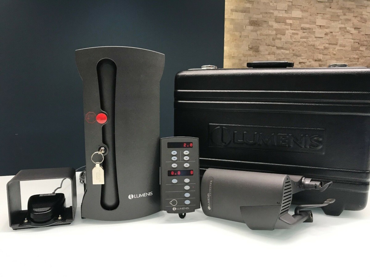
Selective laser trabeculoplasty (SLT) is a procedure that reduces intraocular pressure in patients suffering from glaucoma. The laser is applied using a unique contact lens to the eye’s drainage system, where it stimulates a biochemical change that improves the outflow of fluid from the eye. An SLT laser is one of the lowest powered lasers used in Ophthalmology.
Surgical Microscope
The human eye is a delicate organ, so performing surgery requires monitoring progress on a microscopic level. Surgical microscopes are designed to provide high contrast imaging of all parts of the human eye. When choosing an ophthalmic microscope, it is essential to pay attention to the type of optics employed. An apochromatic lens (or apo) will provide high light transmission, permitting high-quality imaging at lower light intensities. Specific models of ophthalmic surgery microscope provide multiple lighting options, such as switching between halogen and xenon. An ophthalmic surgical microscope can either be fixed or adjustable, and some models offer a second “observer” set of binoculars, some of which can independently adjust the focusing mechanism. Most ophthalmic microscopes are mounted to a rolling stand for versatility and to allow movement around the OR. However, ceiling-mounted microscopes also exist.
Conclusion
Laser Locators specializes in the preventative maintenance and refurbishment of all types of ophthalmic lasers and diagnostics. Whether you are only looking to service an existing device or want to take your practice to the next level, think of us first.
Contact us today for a complimentary consultation on how you can improve your ophthalmic practice.
jo**@la***********.com
by Joey Colarulo, Vice President
About Joey Colarulo
Vice President
Joey has been the Vice President of Laser Locators since March 2015 and a Managing Partner since 2012. He joined the company in 2011.
Joey has significantly contributed to Laser Locators’ growth, including the development of a full service and parts department. He has streamlined the sales and procurement departments by redeveloping processes and implementing new systems. Through Joey’s efforts, Laser Locators has tripled its sales volume and added 13 new positions.
Joey has over 20 years of experience in global internet sales and marketing. His expertise in analyzing the marketplace and leverage the latest e-commerce technologies has enabled Joey to drive exponential sales growth year over year.
Originally from Philadelphia, Joey earned his Bachelor’s degree in Financial Management and graduated Magna Cum Laude from Rowan University.
Outside of work, Joey is involved in the Westchase Charitable Foundation, a local non-profit that provides direct assistance to those in need. His interests include vintage BMWs and rare sports cards.